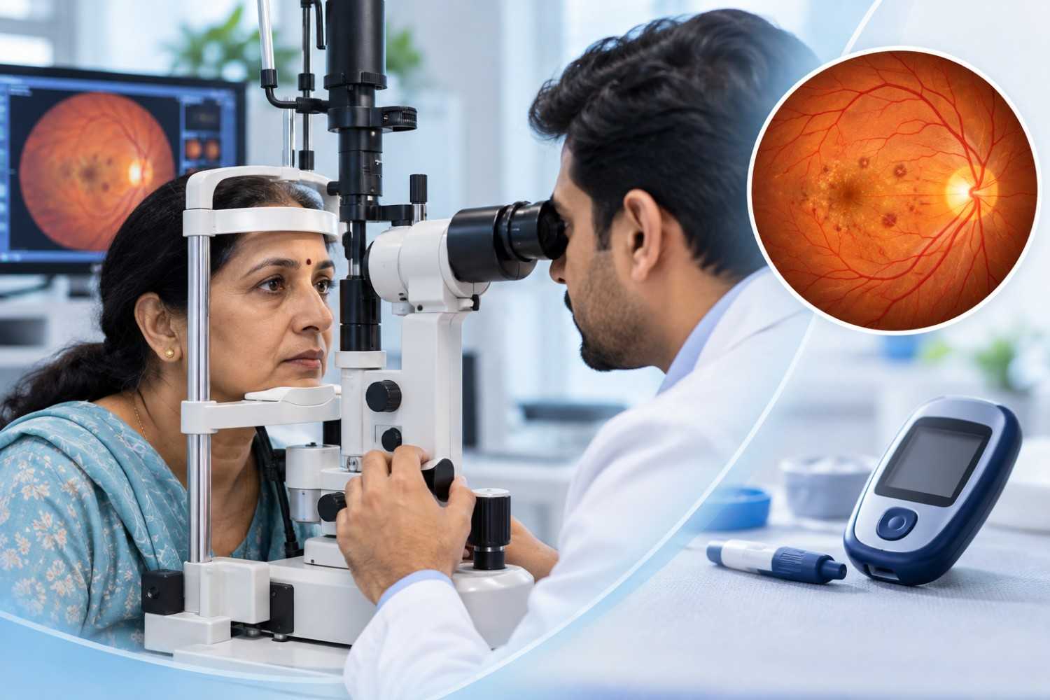
Retina Health for Diabetics in Vadodara: What Regular Screening Involves
Diabetic retinopathy is one of the leading causes of preventable
vision loss in working-age adults, and one of its most frustrating features is
how quietly it progresses. Blood sugar can be damaging the small vessels that
feed the retina for years before a person notices any change in what they see.
India carries a substantial share of the world's diabetes burden, and the
government's National Diabetes and Diabetic Retinopathy Survey found
retinopathy in roughly 17 in every 100 people with diabetes surveyed — a
reminder that this is not a rare complication reserved for advanced cases.
Regular retinal screening is the only practical way to catch these changes
while they are still easy to manage, and this guide explains what that
screening actually involves, how often it is recommended, and what happens if
it turns up something that needs attention.
The Short Answer: What Screening Involves
A diabetic retina screening is a structured eye examination built
specifically to look for damage that diabetes causes at the back of the eye. At
minimum, it includes a visual acuity check, dilation of the pupils with drops,
and a detailed examination of the retina, macula, and optic nerve. Depending on
what is seen, it may also include retinal photography to document findings over
time and optical coherence tomography (OCT) to get a detailed cross-sectional
view of the retina. None of this depends on whether vision currently feels
affected — that is the entire point of screening rather than waiting for
symptoms.
Why Diabetes Affects the Retina
The retina is lined with a dense network of very small blood vessels
that supply the light-sensing tissue with oxygen and nutrients. Chronically
elevated blood sugar damages the walls of these vessels over time, making them
leak fluid and blood into surrounding tissue or, in more advanced stages,
causing the retina to grow fragile new vessels in an attempt to compensate.
Early changes are usually described as non-proliferative diabetic retinopathy —
small vessel damage without new vessel growth — while proliferative diabetic
retinopathy refers to the more advanced stage where abnormal vessels have
formed. Separately, fluid accumulation in the macula, known as diabetic macular
edema, can affect central vision at any stage of retinopathy.
Why It Often Causes No Symptoms Until Late
Retinopathy frequently begins in parts of the retina that are not
responsible for sharp central vision, which means a person can have measurable
disease while still reading comfortably and seeing normally day to day. Vision
usually changes only once the macula becomes involved or once bleeding or
scarring becomes significant — by which point the disease has often had years
to progress. Some people notice fluctuating blur that seems to track with blood
sugar swings and dismiss it for that reason, without realising it can also
reflect retinal changes worth documenting. This gap between disease onset and
noticeable symptoms is exactly why screening is scheduled by time since
diagnosis rather than by how vision feels.
What Happens During a Screening Visit
A typical diabetic retina screening at an eye hospital follows a
consistent sequence:
1.
Visual acuity
testing to establish a baseline reading for each eye.
2.
Dilating eye
drops are applied, which take roughly 15–30 minutes to widen the pupils fully.
3.
A detailed
fundus examination of the retina, macula, blood vessels, and optic nerve using
specialised lenses.
4.
Retinal
photography, when appropriate, to create a documented record that can be
compared at future visits.
5.
OCT imaging,
when indicated, to assess the macula in cross-section and check for fluid or
thickening.
6.
A discussion
of findings and, if needed, a plan for monitoring, treatment, or referral.
Vision typically stays blurred for a few hours after dilation and
bright light can feel uncomfortable, so it is sensible to arrange transport
home or bring sunglasses for the ride back.
How Often Screening Is Recommended
Screening frequency depends mainly on the type of diabetes and how
long it has been present. Guidance from the American Academy of Ophthalmology,
widely reflected in clinical practice, sets out the following starting points:
|
Diabetes Type / Situation |
First Retinal Examination |
Follow-Up (No Retinopathy
Found) |
|
Type 1
diabetes |
Within
5 years of diagnosis |
Annually
thereafter |
|
Type 2
diabetes |
At the
time of diagnosis |
Annually
thereafter |
|
Pregnancy
(pre-existing type 1 or type 2 diabetes) |
Early
in the first trimester |
As
advised by the treating ophthalmologist, often more frequently |
If retinopathy is already present, the treating ophthalmologist will
usually recommend a closer schedule based on its severity, since more advanced
findings need to be monitored more often than the annual baseline.
Risk Factors That Warrant Closer Monitoring
Some factors make diabetic retinopathy more likely to develop or
progress, and they are useful to know even outside a screening appointment:
?
Duration of
diabetes — risk rises the longer diabetes has been
present, regardless of type.
?
Blood sugar
control — higher average blood sugar (HbA1c) over
time is linked to greater retinal risk.
?
Blood
pressure and cholesterol — both can
affect the same small vessels that retinopathy involves.
?
Pregnancy — existing diabetic retinopathy can progress during pregnancy, which
is why earlier and more frequent review is recommended.
?
Kidney
disease — diabetic kidney and eye complications often
track together, since both involve small-vessel damage.
None of these factors guarantee that retinopathy will develop, but
they are reasons a doctor may recommend more frequent screening than the
standard annual interval.
What the Results Might Show
Findings on a diabetic retina screening generally fall into a few
broad categories: no retinopathy, mild-to-moderate non-proliferative changes
such as small vessel dilations or scattered hemorrhages, more advanced
non-proliferative disease, proliferative retinopathy involving abnormal new
vessel growth, and diabetic macular edema affecting central vision. Each
category carries a different recommended monitoring interval and, where
relevant, a different treatment discussion. A finding of mild retinopathy is not
a diagnosis of ongoing vision loss — it is information that supports earlier
intervention, which is generally more effective than treatment started after
the disease has progressed further.
What Happens If Changes Are Found
When a screening identifies diabetic retinopathy or macular edema, the
next step depends on severity. Mild changes are often monitored with more
frequent follow-up examinations while blood sugar, blood pressure, and
cholesterol management continue with the treating physician. More significant
changes may call for anti-VEGF injections to reduce abnormal vessel activity
and macular swelling, laser photocoagulation to stabilise areas of the retina
at risk, or, in advanced cases involving bleeding or scar tissue, vitrectomy
surgery to clear the vitreous and repair the retina. Which option applies, if
any, depends entirely on what the examination and imaging show for that
specific eye — there is no single treatment path that fits every diagnosis, and
a treating ophthalmologist is best placed to explain the reasoning for a
particular case.
FAQ
Can diabetic retinopathy
occur without any symptoms?
Yes. Early diabetic retinopathy very often causes no noticeable change
in vision, which is exactly why screening is scheduled by time since diagnosis
rather than by how the eyes feel.
How often should a person
with diabetes have an eye examination?
As a general starting point, people with type 1 diabetes are typically
screened within 5 years of diagnosis and annually after that, while people with
type 2 diabetes are usually screened at diagnosis and annually thereafter.
Pregnancy and existing retinal findings can shorten this interval, so the
treating ophthalmologist should confirm the right schedule for each individual.
Does good blood sugar
control mean I don't need screening?
No. Good control lowers the risk of retinopathy developing or
progressing, but it does not eliminate the need for screening, since retinal
changes can still occur and are only reliably detected through examination.
Is dilation necessary for
a diabetic eye screening?
Dilating drops allow a much wider and more thorough view of the retina
than an undilated exam, which is why dilation is considered a standard part of
an adequate diabetic retina screening.
Can diabetic retinopathy
be treated if caught early?
Early and moderate changes are generally more straightforward to
monitor and manage than advanced disease, which is the main argument for
regular screening rather than waiting for symptoms. Specific treatment
recommendations depend on the findings in each case.
Will my vision stay
blurry after the screening appointment?
Yes, temporarily. Dilating drops typically blur near vision and
increase light sensitivity for a few hours, so many patients prefer to arrange
a ride home and bring sunglasses for the journey.