Fundus & Anterior Segment Photography in Vadodara

Fundus and Anterior Segment Photography at Sattva Eye Hospital Vadodara
Fundus Photography in Vadodara

Detailed Fundus & Anterior Segment Photography at Sattva Eye Hospital

A lot of eye problems don't hurt until real damage has already been done. Diabetic retinopathy, glaucoma, and early macular changes can sit quietly at the back of your eye for months without a single symptom. That is exactly what fundus photography is built to catch.

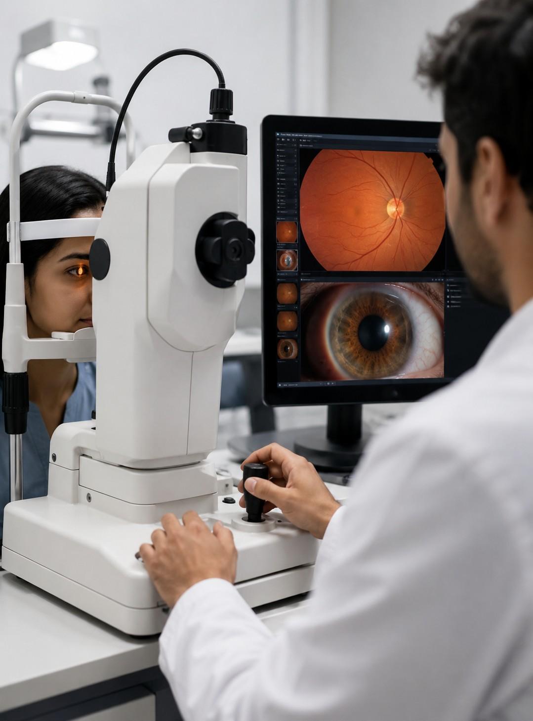
At Sattva Eye Hospital in Vadodara, we use digital fundus and anterior segment photography to capture a clear, detailed image of your retina, optic nerve, cornea, and other eye structures, giving your ophthalmologist something precise to work with instead of a guess based on a quick look. The test takes only a few minutes, causes no discomfort, and gives you a permanent visual record you can compare against on every future visit.

About the Service: Fundus photography photographs the back of your eye, including the retina, optic disc, macula, and the blood vessels that run through it. Anterior segment photography does the opposite job, documenting the front structures such as the cornea, iris, lens, and surrounding tissue.

Together, the two give a complete visual record of your eye that goes well beyond what a routine exam can capture on its own. The images also help your ophthalmologist identify and monitor changes more accurately over time.

This test is useful for people with diabetes or high blood pressure who need regular retina monitoring, anyone with a family history of glaucoma or macular degeneration, patients being tracked before or after eye surgery, and anyone who wants a proper baseline record of their eye health.

Fundus and anterior segment photography also supports a comprehensive eye examination by giving the doctor objective images to refer back to, rather than relying only on notes or memory from one visit to the next.

Book Your Fundus Photography Appointment in Vadodara — Call Sattva Eye Hospital or schedule your appointment online. Most reports are ready the same day.

Why Choose Sattva Eye Hospital for Fundus Photography in Vadodara

Experienced ophthalmology team with years of hands-on diagnostic and surgical practice
Advanced digital fundus and anterior segment camera for sharp, high-resolution imaging
Quick, non-invasive test that fits into a single visit with no downtime
Every image is reviewed personally by a consulting ophthalmologist, not just a technician
Comfortable, patient-friendly setup with minimal waiting time
Transparent, upfront pricing with no hidden charges
Convenient Vadodara location with easy scheduling for walk-ins and appointments
Fundus Photography

Our Fundus Photography Process

Our fundus and anterior segment photography process is simple, comfortable, and designed to give your ophthalmologist clear images for accurate evaluation and timely guidance.

Consultation & History

We start with a short conversation about your symptoms, medical history, and existing conditions such as diabetes or hypertension that may affect your eye health.

Pupil Dilation, If Needed

For a wider view of the retina, we may use dilating drops. These usually take around fifteen to twenty minutes to take effect.

Imaging

You rest your chin on the camera stand while our technician captures fundus and anterior segment images. There is no contact with your eye and no pain involved.

Doctor Review

A consulting ophthalmologist examines the images the same day and carefully looks for any signs or changes that may need further attention.

Report & Explanation

We walk you through the findings in plain language and provide a printed or digital copy of your report for your records and future reference.

Follow-Up Plan

If anything needs further attention, we discuss the next steps with you right away, so you are not left waiting for a callback or wondering what to do next.

FUNDUS & ANTERIOR SEGMENT PHOTOGRAPHY

What's Included in Our Fundus Photography Service

Our digital eye photography service provides detailed images of the retina and front structures of the eye, supported by expert ophthalmologist review and clear guidance for further care when required.

Digital Fundus Imaging

High-resolution digital fundus imaging covering the retina, optic nerve, and macula to provide a clear visual record of the back of the eye.

Anterior Segment Imaging

Detailed imaging of the front structures of the eye, including the cornea, iris, and lens, for accurate documentation and clinical assessment.

Image Comparison

Where previous scans are available, we compare images over time to help identify and track changes in the condition of your eyes.

Ophthalmologist Review

Every set of images is reviewed by a consulting ophthalmologist, with findings documented in a clear written report.

Clear Next-Step Guidance

Based on the findings, we provide clear guidance about the next steps, treatment options, monitoring, or referral to another specialist when required.

Benefits of Choosing Our Fundus Photography Service

Catches silent conditions like diabetic retinopathy before symptoms show up
Builds a baseline record you can track against for years to come
Cuts down on repeated manual exams by giving doctors a clear image to reference
Completely non-invasive, with no needles and no discomfort
Digital images that can be shared easily if you ever need a second opinion
Supports a more proactive, rather than reactive, approach to eye health

Frequently Asked Questions

Find answers to common questions about fundus photography, anterior segment photography, eye imaging, preparation, procedure time, and fundus photography at Sattva Eye Hospital, Vadodara.

Fundus photography is a diagnostic test that takes a detailed photograph of the back of your eye, including the retina, optic nerve, and macula. It helps your doctor spot changes or damage that aren't visible during a standard eye exam.

No. The test is completely non-invasive. You simply rest your chin on a stand while the camera captures the image. There's no contact with the eye itself and no pain involved.

Dilation isn't always required, but it's often recommended for a wider, clearer view of the retina. If needed, we'll apply dilating drops that take about fifteen to twenty minutes to work.

The imaging itself takes just a few minutes. With consultation, dilation, and report review included, most visits are wrapped up within an hour.

Fundus photography captures the back of the eye, including the retina, optic nerve, and macula. Anterior segment photography captures the front, including the cornea, iris, and lens. Together they give a complete picture of your eye health.

It's especially useful for people with diabetes or high blood pressure, anyone with a family history of glaucoma or macular degeneration, patients being monitored before or after eye surgery, and anyone who simply wants a baseline record of their eye health.

For most people with diabetes or hypertension, an annual scan is recommended. Your ophthalmologist may suggest a different frequency depending on your specific condition.

Pricing depends on whether dilation and additional imaging are required. Contact our Vadodara clinic directly for current pricing and to book an appointment.