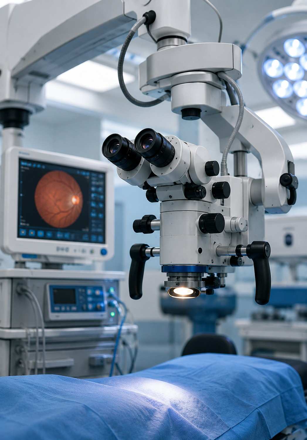
Cataract removal, retina repair, corneal grafts, glaucoma procedures — different as these surgeries are, they all happen under the same essential piece of equipment: the ophthalmic operating microscope. It doesn't cut, suction or implant anything itself, but it plays a critical role in surgery because every instrument is guided by what the surgeon can actually see through it.
At Sattva Eye Hospital in Vadodara, an advanced ophthalmic microscope is used during our eye surgeries, from routine cataract procedures to more complex vitreoretinal work. It provides the magnification, illumination and depth perception required for precise surgical visualization.
What an Ophthalmic Microscope Does: Unlike a regular laboratory microscope that magnifies a flat, static sample, an ophthalmic surgical microscope is designed to provide a magnified, three-dimensional view of a living eye. It maintains a suitable working distance so the surgeon can comfortably operate with surgical instruments under the microscope.
The microscope uses two separate eyepieces positioned at a binocular angle. The surgeon's brain combines these two views to create genuine depth perception, known as stereopsis. This stereo visualization helps the surgeon understand the position and depth of delicate eye structures during surgery.
Red Reflex Illumination: During cataract surgery, the microscope's illumination can be aligned closely with the surgeon's viewing axis. Light passes through the pupil, reflects from the retina and returns as a reddish-orange glow known as the red reflex.
This red reflex helps the surgeon clearly distinguish the cloudy lens and remaining lens fragments against the illuminated background. Maintaining a stable and consistent red reflex, even when the patient's eye moves slightly, is an important part of effective ophthalmic surgical visualization.
Experience Advanced Eye Surgery Technology in Vadodara — Consult the ophthalmology team at Sattva Eye Hospital to learn more about microscope-assisted eye surgery and advanced surgical visualization.
Modern ophthalmic microscopes are designed not only for precise eye surgery visualization, but also to support surgeon comfort and carefully controlled illumination throughout the procedure.
Modern ophthalmic microscopes allow surgeons to maintain a comfortable and stable working posture, which is especially important during procedures that may take longer to complete.
Adjustable eyepiece angles allow the surgeon to position the viewing system according to their working posture, helping maintain comfortable visualization throughout surgery.
Balanced construction and motorised positioning help reduce unnecessary physical strain, allowing the surgeon to remain steady and focused during delicate eye surgery.
A stable ergonomic setup can help minimise fatigue during lengthy procedures, supporting consistent hand control and precise surgical movements.
Modern surgical microscopes provide adjustable illumination, allowing the surgical team to control the amount of light used according to the requirements of each surgical step.
Built-in filters and intensity controls help the surgical team adjust or reduce illumination during less critical moments instead of continuously operating at maximum brightness.
Modern ophthalmic microscopes can integrate digital displays and advanced imaging systems, helping the surgical team view magnified details and correlate pre-operative scans with the live surgical view.
Advanced microscope systems can transmit the surgical view to a large operating theatre screen in real time, allowing the surgical team to follow the procedure clearly.
A larger display allows the surgical team to follow magnified anatomical details while the surgeon can cross-check the microscope view against the wider screen when required.
Some advanced systems can integrate diagnostic imaging captured before surgery with the microscope's live surgical view, supporting more informed visualization.
Real-time screen sharing can help other members of the surgical team follow the procedure without needing to look directly through the microscope eyepieces.
In selected retina and corneal procedures, matching pre-surgical diagnostic scans with the live surgical view can provide an additional layer of anatomical reference.
When choosing an eye hospital in Vadodara, patients can ask whether the microscope is dedicated to ophthalmic surgery, regularly serviced, and equipped with the illumination and viewing attachments required for their specific procedure.
Find answers to common questions about ophthalmic microscopes, red reflex illumination, surgical visualization, microscope safety, and eye surgery at Sattva Eye Hospital, Vadodara.