Ultrasound A-Scan & B-Scan Testing in Vadodara

A-Scan and B-Scan Ultrasound Test in Vadodara at Sattva Eye Hospital
A-Scan & B-Scan Ultrasound Test in Vadodara

A-Scan & B-Scan Ultrasound for Detailed Eye Examination

Most eye tests rely on light, whether that's a camera photographing the retina or an OCT scanner mapping it in cross-section. Ultrasound testing works differently: it uses sound waves, which is exactly why it's useful in situations where light can't get through, and why it remains one of the more dependable tools in an eye clinic even in the age of high-resolution imaging.

At Sattva Eye Hospital in Vadodara, we use two related tests built on this principle: the A-scan, mainly to measure the eye before cataract surgery, and the B-scan, mainly to look inside the eye when something else is blocking the view.

What A-Scan and B-Scan Actually Measure

An A-scan, short for amplitude scan, sends a single beam of sound through the eye and records it as a line of spikes, each one marking a boundary between different internal structures. Its main job is measuring the eye's axial length, the distance from front to back, which is one of the key numbers used to calculate the correct power of the intraocular lens (IOL) for cataract surgery.

A B-scan, or brightness scan, builds a full two-dimensional cross-sectional picture of the eye's interior instead of a single line, similar in principle to how an ultrasound builds a picture rather than a single measurement. That makes it the test of choice when a doctor needs to actually see the shape and structure of the retina, vitreous or surrounding tissue, not just measure a distance.

Book Your A-Scan & B-Scan Ultrasound Test in Vadodara — Contact Sattva Eye Hospital for an eye ultrasound evaluation and expert ophthalmic assessment.

Why We Use A-Scan & B-Scan Ultrasound at Sattva Eye Hospital

A-scan accurately measures the eye's axial length, an essential measurement for calculating the correct IOL power before cataract surgery
A-scan may be combined with keratometry to provide the measurements needed for personalised IOL power calculation
B-scan provides a two-dimensional view of the posterior segment when the retina cannot be seen directly
Helps detect retinal detachment hidden behind a dense cataract or vitreous haemorrhage
Helps assess bleeding inside the eye following trauma or advanced diabetic retinopathy
Helps identify suspected intraocular tumours, masses or other abnormal structures inside the eye
Helps detect retained foreign material and structural changes at the back of the eye, including in high myopia
B-scan can provide important information before vitrectomy or other retinal surgery when blood or cloudiness blocks the normal view
A-Scan & B-Scan Ultrasound

What Happens During the Test

A-scan and B-scan ultrasound tests are performed in the clinic, usually take only a few minutes, and do not involve any radiation. The procedure used depends on whether the doctor needs to measure the eye or examine its internal structures.

A-Scan Preparation

For an A-scan, a numbing drop is placed in the eye before the examination. This helps make the measurement comfortable while the ultrasound probe is positioned carefully.

A-Scan Measurement

A small ultrasound probe gently touches the surface of the eye to measure its axial length. The test is quick and provides an important measurement for IOL power calculation.

B-Scan Ultrasound

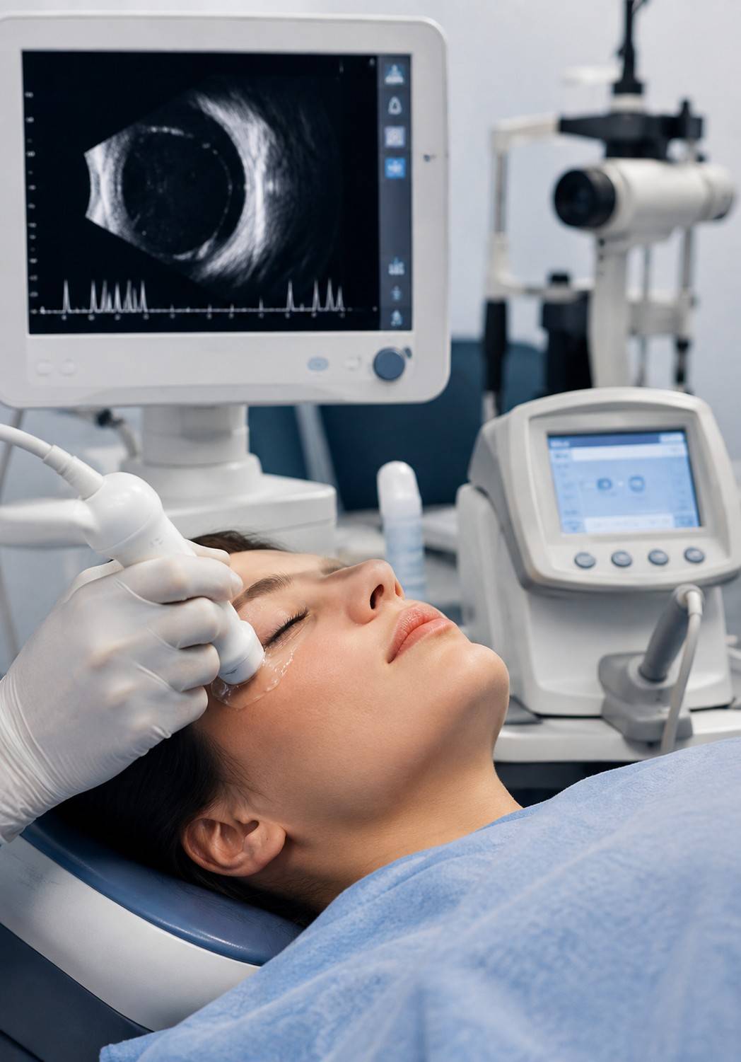
B-scan is generally performed through the closed eyelid using a small amount of ultrasound gel. It does not require direct contact with the surface of the eye.

Quick & Radiation-Free

Both ultrasound examinations are performed in the clinic, usually take only a few minutes, and use sound waves rather than radiation. Patients can blink and move normally between readings.

Optical Biometry Alternative

Optical biometry is a light-based alternative that measures axial length without touching the eye and is often used first for routine cataract cases when the optical path is sufficiently clear.

When A-Scan Is Preferred

A-scan remains a dependable option when a very dense cataract interferes with optical biometry. Keeping both technologies available allows the appropriate measurement method to be selected for each patient.

A-SCAN & B-SCAN ULTRASOUND

Who Typically Needs This Test

A-scan and B-scan ultrasound can be recommended in different situations, from routine cataract surgery planning to evaluating the inside of the eye when the retina cannot be viewed clearly.

Cataract Surgery Planning

Anyone being evaluated for cataract surgery may need eye biometry as part of routine pre-surgical planning, helping the surgeon determine the appropriate IOL power.

Dense Cataract

Patients with a very dense cataract that blocks a clear view of the retina may need B-scan ultrasound to assess the structures behind the cloudy lens.

Vitreous Haemorrhage

Patients with vitreous haemorrhage, particularly when associated with advanced diabetic eye disease, may need B-scan imaging when blood prevents direct examination of the retina.

Recent Eye Trauma

Anyone with recent eye trauma may require ultrasound evaluation when the doctor needs to check internal eye structures or assess possible damage that cannot be seen clearly from the outside.

Suspected Eye Mass

Patients being evaluated for a suspected mass or growth inside the eye may benefit from B-scan ultrasound to examine the shape and internal structures of the eye.

Choosing a Centre for Ultrasound Eye Testing in Vadodara

B-scan is an interpretive examination, so experience in performing and reviewing ophthalmic ultrasound can be important for accurate assessment
Retina-focused assessment can help ensure that posterior segment findings are interpreted in the right clinical context
Both A-scan and B-scan ultrasound are available on-site for convenient diagnostic and pre-surgical evaluation
Pre-surgical eye measurements and diagnostic ultrasound can often be completed during the same visit
Useful for both routine cataract planning and situations where the retina or posterior segment cannot be viewed directly
On-site availability helps reduce delays when an ultrasound assessment is needed before deciding on further treatment or surgery

Frequently Asked Questions

Find answers to common questions about A-scan and B-scan ultrasound testing, eye biometry, cataract surgery planning, retinal imaging, test duration, radiation, and results at Sattva Eye Hospital, Vadodara.

No. An A-scan involves a brief, gentle touch on a numbed eye, while a B-scan is usually done through the closed eyelid with gel, similar to an external ultrasound. Neither test is generally described as painful.

Both A-scan and B-scan tests are typically completed within a few minutes per eye. Your overall visit may take longer if other pre-surgical or diagnostic checks are performed at the same time.

Yes. Some form of eye biometry, whether ultrasound A-scan or optical biometry, is a standard part of planning the correct intraocular lens (IOL) power for cataract surgery. Your ophthalmologist will recommend the appropriate method based on your eye.

In many cases, yes. B-scan ultrasound can help image the position and structure of the retina even when a dense cataract or bleeding blocks the direct view normally used by your doctor. The findings are interpreted along with your clinical examination.

No. Both A-scan and B-scan ultrasound use sound waves rather than radiation. They use the same underlying ultrasound principle as other diagnostic ultrasound examinations.

Yes. Both tests can be reviewed and reported during your visit. Your ophthalmologist will discuss the findings with you and explain any recommended next steps based on the results.